
Introduction
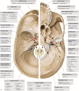
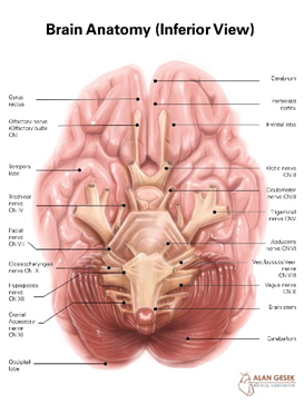
Skull base surgery is one of the most complex and delicate fields in modern neurosurgery. The skull base forms the floor of the brain and separates it from critical structures such as the eyes, nasal cavity, sinuses, major blood vessels, and cranial nerves. Tumors and lesions in this region are particularly challenging because of their proximity to vital neurological and vascular structures.
Advances in microsurgical and endoscopic techniques have revolutionized skull base surgery, allowing surgeons to treat previously inoperable conditions with greater safety, precision, and improved patient outcomes.
Understanding the Skull Base
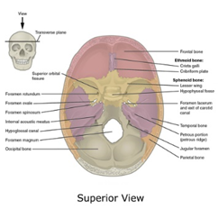
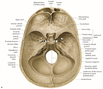
The skull base is anatomically divided into three regions:
- Anterior skull base (near the frontal lobes and sinuses)
- Middle skull base (housing the pituitary gland, optic nerves, and carotid arteries)
- Posterior skull base (near the brainstem and cranial nerves)
Lesions in these areas may cause visual problems, hormonal disorders, facial numbness, hearing loss, or balance disturbances.
Conditions Treated with Skull Base Surgery
Skull base surgeries are performed to treat a wide range of conditions, including:
- Pituitary adenomas
- Meningiomas
- Chordomas and chondrosarcomas
- Acoustic neuromas (vestibular schwannomas)
- Craniopharyngiomas
- Cerebrospinal fluid (CSF) leaks
- Vascular lesions involving the skull base
Each condition requires a carefully tailored surgical approach.
Pituitary Surgery
The Pituitary Gland
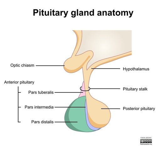
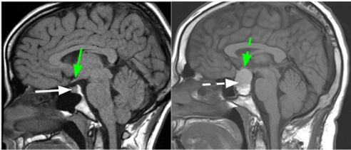
The pituitary gland is a small but vital structure located at the skull base, responsible for regulating many of the body’s hormonal functions. Tumors of the pituitary gland may cause hormonal imbalances, vision loss, headaches, and fatigue.
Surgical Approaches
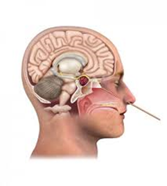
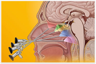
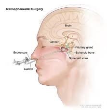
Modern pituitary surgery is most commonly performed using a minimally invasive endoscopic transsphenoidal approach, which allows surgeons to reach the pituitary gland through the nasal passages without external incisions.
Advantages of endoscopic pituitary surgery include:
- Improved visualization of the tumor
- Minimal disruption to surrounding brain tissue
- Reduced postoperative pain
- Faster recovery and shorter hospital stay
- Better preservation of hormonal and visual function
Surgical Approaches to Skull Base Lesions
1. Endoscopic Skull Base Surgery
- Performed through the nose or natural openings
- Ideal for midline skull base and pituitary lesions
- Minimizes brain retraction and cosmetic impact
2. Microsurgical Skull Base Surgery
- Uses high-powered operating microscopes
- Necessary for large, complex, or laterally located tumors
- Allows precise dissection around cranial nerves and blood vessels
3. Combined Approaches
In complex cases, endoscopic and microsurgical techniques may be combined to maximize tumor removal while preserving neurological function.
Advanced Technologies in Skull Base Surgery
State-of-the-art skull base surgery relies on:
- Neuronavigation systems for real-time anatomical guidance
- Intraoperative neuro-monitoring to protect cranial nerves
- High-definition endoscopes and microscopes
- Advanced imaging (MRI & CT fusion) for precise planning
These technologies significantly improve surgical accuracy and safety.
Postoperative Care and Recovery
After skull base surgery, patients receive specialized care including:
- Neurological and hormonal monitoring
- Visual assessment when optic nerves are involved
- Management of CSF leaks or nasal healing
- Follow-up imaging to assess surgical outcomes
Most patients experience significant symptom improvement, especially when surgery is performed early.
Risks and Outcomes
While skull base surgery is highly complex, modern techniques have greatly reduced risks. Outcomes depend on:
- Tumor type and size
- Location and involvement of critical structures
- Patient’s neurological condition before surgery
- Surgical expertise and technology used
When performed by experienced teams, skull base surgery offers excellent long-term control and quality of life improvement.