
Precision, Safety, and Faster Recovery in Modern Brain & Spine Surgery
Introduction
Minimally invasive neurosurgery (MIN) represents a major evolution in the surgical treatment of disorders affecting the brain, spine, spinal cord, and peripheral nerves. Unlike traditional open surgery, minimally invasive techniques aim to achieve the same or better surgical results through smaller incisions, less tissue disruption, and greater precision.
These advanced techniques reduce surgical trauma, shorten hospital stays, accelerate recovery, and minimize postoperative complications—while maintaining the highest standards of safety and effectiveness.
What Is Minimally Invasive Neurosurgery?
Minimally invasive neurosurgery uses specialized instruments, operating microscopes, endoscopes, neuronavigation systems, and real-time imaging to access deep or delicate neurological structures through small, strategically placed openings.
Rather than large incisions and extensive tissue exposure, surgeons work through:
- Small skin incisions
- Natural anatomical corridors
- Keyhole approaches
- Endoscopic pathways
This approach preserves healthy tissue and reduces the physiological stress of surgery.
Minimally invasive neurosurgery (MIN) represents the modern conceptual basis of the new centuries neurosurgical practice. In MIN the target pathology is addressed and surgically treated utilizing the new technological devices like the surgical microscope, the endoscopes or image guiding machines to minimize and sometimes operate without any incisions as in Stereotactic Radio Surgery (SRS).
Minimally Invasive neurosurgery have modified the neurosurgeons perception of the patient’s problem and has lead to minimizing the surgical incisions when performing key hole surgeries where natural corridors are utilized to approach specific areas.
The surgical microscope constitutes the major corner stone in the field of MIN which may be used individually or assisted endoscopically or even using image guidance to ensure accurate and efficient achievement of the surgical goal.
Minimally invasive neurosurgery has revolutionary altered the outcome in the fields of neuro-oncology, pediatric neurosurgery, functional neurosurgery and spinal cord and column surgeries.
Much smaller incisions like the eye brow or small straight incisions to perform mini craniotomies or the utilization of the nasal nostril have markedly minimized tissue disruption, lead to satisfying cosmetic outcome and significantly reduced the hospital stay and incidence of infection and thus had a positive economic result as well.
Key Principles of Minimally Invasive Neurosurgery
- Maximum precision, minimal disruption
- Preservation of neurological function
- Reduced pain and blood loss
- Faster functional recovery
- Improved cosmetic outcomes
Conditions Treated with Minimally Invasive Neurosurgery
1. Brain Tumors
Many brain tumors can now be treated using minimally invasive approaches, including:
- Meningiomas
- Pituitary adenomas
- Selected gliomas
- Skull base tumors
Techniques such as keyhole craniotomy and endoscopic tumor removal allow precise access while minimizing brain retraction.
2. Skull Base & Pituitary Disorders
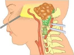
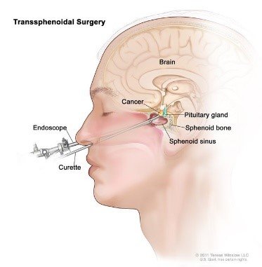
Endoscopic skull base surgery enables surgeons to reach tumors through the nasal passages, avoiding external incisions.
Benefits include:
- No visible scars
- Less postoperative pain
- Shorter hospital stay
- Improved visual and hormonal outcomes
3. Minimally Invasive Spine Surgery
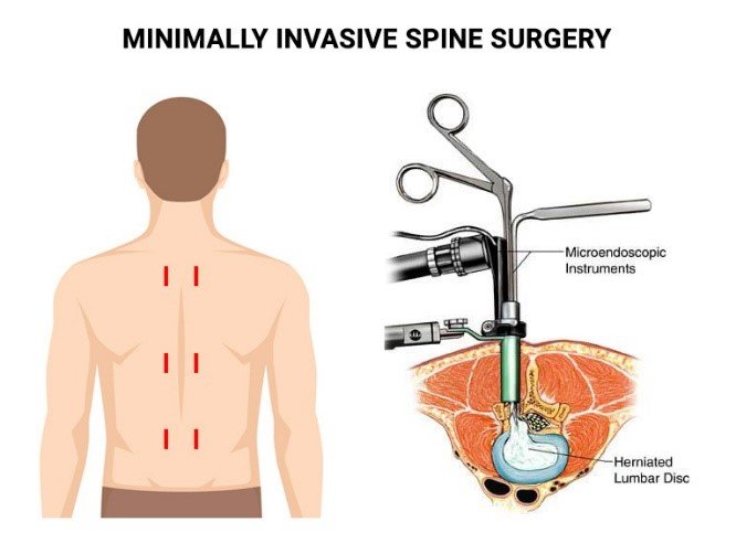
Minimally invasive spine surgery (MISS) is widely used for:
- Herniated discs
- Spinal stenosis
- Degenerative spine disease
- Selected spinal fractures
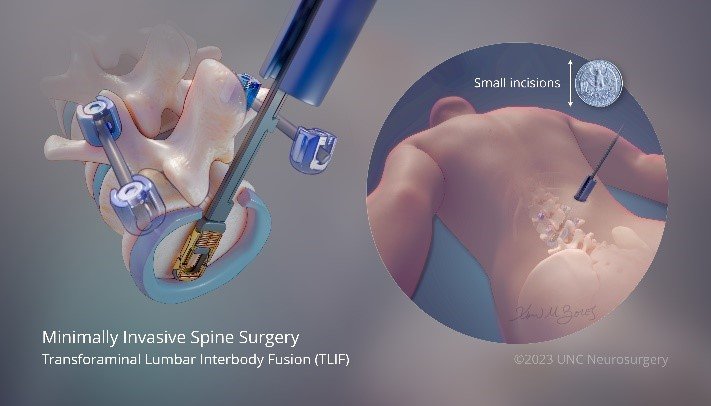
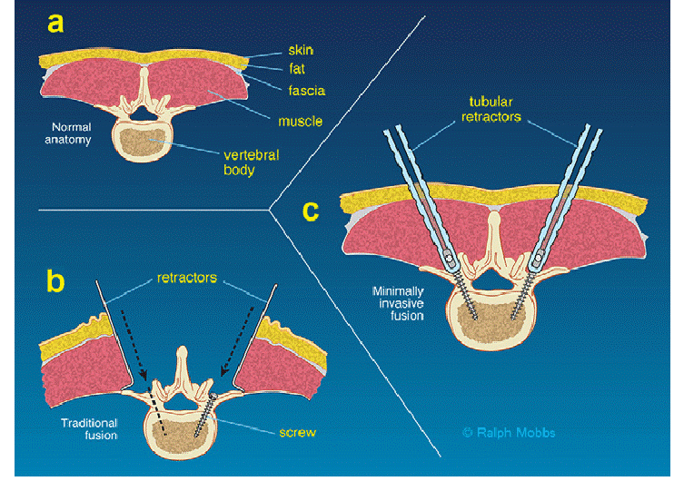
These procedures use tubular retractors, endoscopes, and percutaneous fixation techniques to protect muscles and ligaments.
4. Vascular Neurosurgery
Selected vascular conditions, such as aneurysms and AVMs, can be managed using minimally invasive or endovascular techniques, reducing surgical risk while maintaining efficacy.
Advanced Technologies Enabling Minimally Invasive Neurosurgery
Modern minimally invasive neurosurgery relies on:
- Neuronavigation systems for real-time anatomical guidance
- High-definition operating microscopes
- Endoscopic visualization
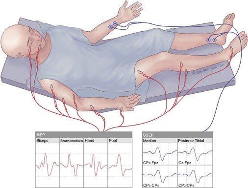
- Intraoperative neuro-monitoring to protect motor and sensory pathways
- Advanced MRI and CT imaging integration
These tools allow surgeons to operate with millimeter-level accuracy.
Micro-Neurosurgery
Surgical Microscopes provide Magnification and Illumination for neurosurgeon allowing them to perform precisely and efficiently their tasks via narrow and deep corridors. Specially designed microsurgical instruments are utilized in performing neurological or spinal surgical procedures. Micro-neurosurgery have modified the morbid surgical outcome into a more tolerable and easy recovery for patients who are suffering several problems including, Neuro-Oncology, Vascular Neurosurgery, Pediatric Neurosurgery, Spinal Surgery and Functional Neurosurgery.
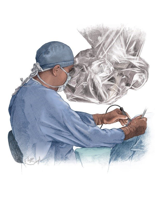
Micro-neurosurgical procedures can either be performed using the surgical microscope alone or is assisted with neuro-endoscopes or sometimes intraoperative neuro-navigative and image guidance to ensure complete and accurate achievement of the intended goals for surgery. Our operating room is equipped with a Carl Zeiss surgical microscope model ——–, which offers a floating maneuverability during surgery and facilitates for an assistant surgeon to participate via the side viewer and also the other operating room members to observe via the on screen transmission from the microscope field.
Micro-neurosurgery constitutes a cornerstone in the field of Minimally Invasive Neurosurgery and its role in performing Key hole surgeries via small skin incisions and small skull window craniotomies in treating complex tumors or vascular pathologies can’t be undertake. Spinal disc diseases are in many instances approached microscopically; anterior cervical discectomy and lumbar posterior micro-discectomy. Furthermore, the surgical microscope is mandatory in spinal cord tumors, Vascular aneurysms and vascular malformations are all managed micro-surgically.
Neuro-Endoscopy
With the advancement of fiber-optics and the development of surgical endoscopes, many neurosurgical procedures were modified to become much easier and less invasive to the patients and with even much better outcomes.
The Neuro-Endoscope also provides magnification and illumination to the surgeon like the surgical microscopes. Our Olympus model camera, Olympus model light source and digital recording system, all facilitate for us to perform complex operations.
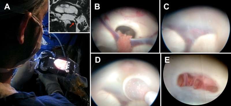
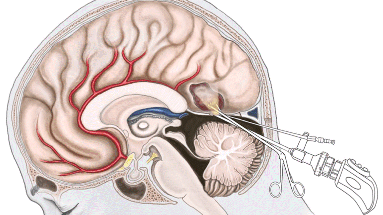
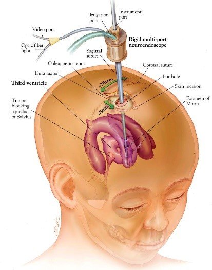
The neuro-endoscope has helped a lot in diseases related to the cerebrospinal fluid (CSF) dynamics owing to the development of wide CSF filled spaces, including Hydrocephalus, multi-loculated/compartmental hydrocephalus and arachnoid cysts. Intraventricular lesions in general and 3rd ventricular colloid cysts in particular are quite efficiently managed endoscopically. Pituitary gland tumors are increasingly being operated endoscopically through the patient’s nostril via the skull base without any incisions. Also the endoscope is used in the extended trans-nasal trans-basal approaches that allow for resection of skull base tumors like meningiomas and craniopharyngiomas and unfortunately repairs of CSF leaks from the anterior skull base. Spinal endoscopy is quite limited, yet lumbar disc surgeries may be performed endoscopically. Anterior trans-thoracic or trans- abdominal laparoscopic approaches to the vertebral column discs are sometimes indicated procedures.
The neuro-endoscopes may be used in assistance to the surgical microscope to ensure complete resection of tumors especially around hidden corners and in microvascular decompression in Trigeminal neuralgias to explore the nerve’s ventral aspect.
The neuro-endoscope constitutes an essential instrument intra-operatively that can perform both diagnostic and therapeutic procedures.
Image Guided Neurosurgery
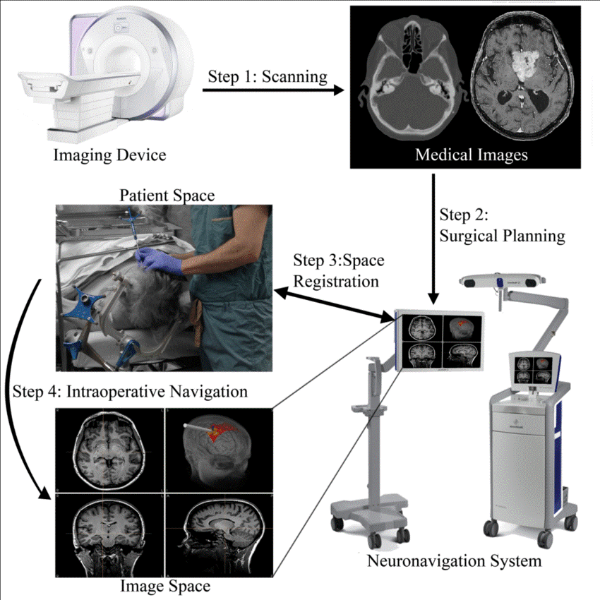
Frameless Image guided surgery (IGS) in neurosurgery is the technology where the patients radiological images whether computed tomography (CT)-scan, magnetic resonance imaging (MRI) or plan x-rays are transmitted to the operating theater on a special machine and the patient is registered to these machines in a three dimension reconstruction, together with the surgical instruments to provide real time motion of the surgical instruments in relation to the intended pathology with accuracies of up to 0.4 mm. This technology facilitates the resection of tumors in eloquent areas like the motor or speech areas and also deeply seated ones especially those with indefinite outlines. The use of IGS in neurosurgery also plays a role in spinal instrumentation where the length and direction of the desired pedicular screw are easily determined, thus minimizing the incidence of complications related to system failures.
Stereotactic fixed frame based systems allow for precise shunt placement, biopsy taking, electrode placement in Deep Brain Stimulation (DBS) in the treatment of Parkinson’s disease.
The CNS center operating room is equipped with an up to date Neuro-navigation device, the brain Lab model ——-, that reconstruct’s CT/ MRI/ or X-ray pictures of the patient allowing for more accurate performance during the operation from the decision of the skin incision till the end of the operation.
Advantages for Patients
Compared to traditional open surgery, minimally invasive neurosurgery offers:
- Smaller incisions and less scarring
- Reduced postoperative pain
- Lower risk of infection and blood loss
- Shorter hospital stays
- Faster return to daily activities
- Improved overall patient experience
These benefits are especially important for elderly patients and those with complex medical conditions.
Postoperative Care and Recovery
Recovery after minimally invasive neurosurgery is typically smoother and faster. Postoperative care includes:
- Early mobilization
- Pain control with reduced need for medications
- Shorter intensive care monitoring
- Early rehabilitation when required
Most patients can resume normal activities sooner than with traditional surgery.
Limitations and Patient Selection
Not all conditions are suitable for minimally invasive approaches. Careful patient selection is essential and depends on:
- Tumor size and location
- Vascular involvement
- Patient’s overall health
- Surgeon’s experience and available technology
When appropriately selected, outcomes are equal or superior to conventional surgery.
The Role of Experience and Expertise
While technology plays a vital role, surgical expertise remains the most important factor in successful minimally invasive neurosurgery. Mastery of anatomy, precise planning, and extensive experience are critical to achieving safe and effective results.
Future of Minimally Invasive Neurosurgery
Ongoing advances in:
- Robotics
- Augmented reality
- Artificial intelligence
- Real-time imaging
are expected to further expand the capabilities and safety of minimally invasive neurosurgical procedures.
When to Consider Minimally Invasive Neurosurgery
Patients should seek specialist evaluation if they:
- Require brain or spine surgery
- Wish to explore less invasive treatment options
- Are concerned about recovery time or cosmetic outcomes
- Have been advised surgery after failed conservative treatment
A specialized neurosurgical consultation can determine the most appropriate approach.
Conclusion
Minimally invasive neurosurgery has transformed the way complex neurological disorders are treated. By combining advanced technology, refined surgical techniques, and deep anatomical knowledge, surgeons can now achieve excellent outcomes with less trauma and faster recovery.
The ultimate goal is to treat disease effectively while preserving function, dignity, and quality of life.