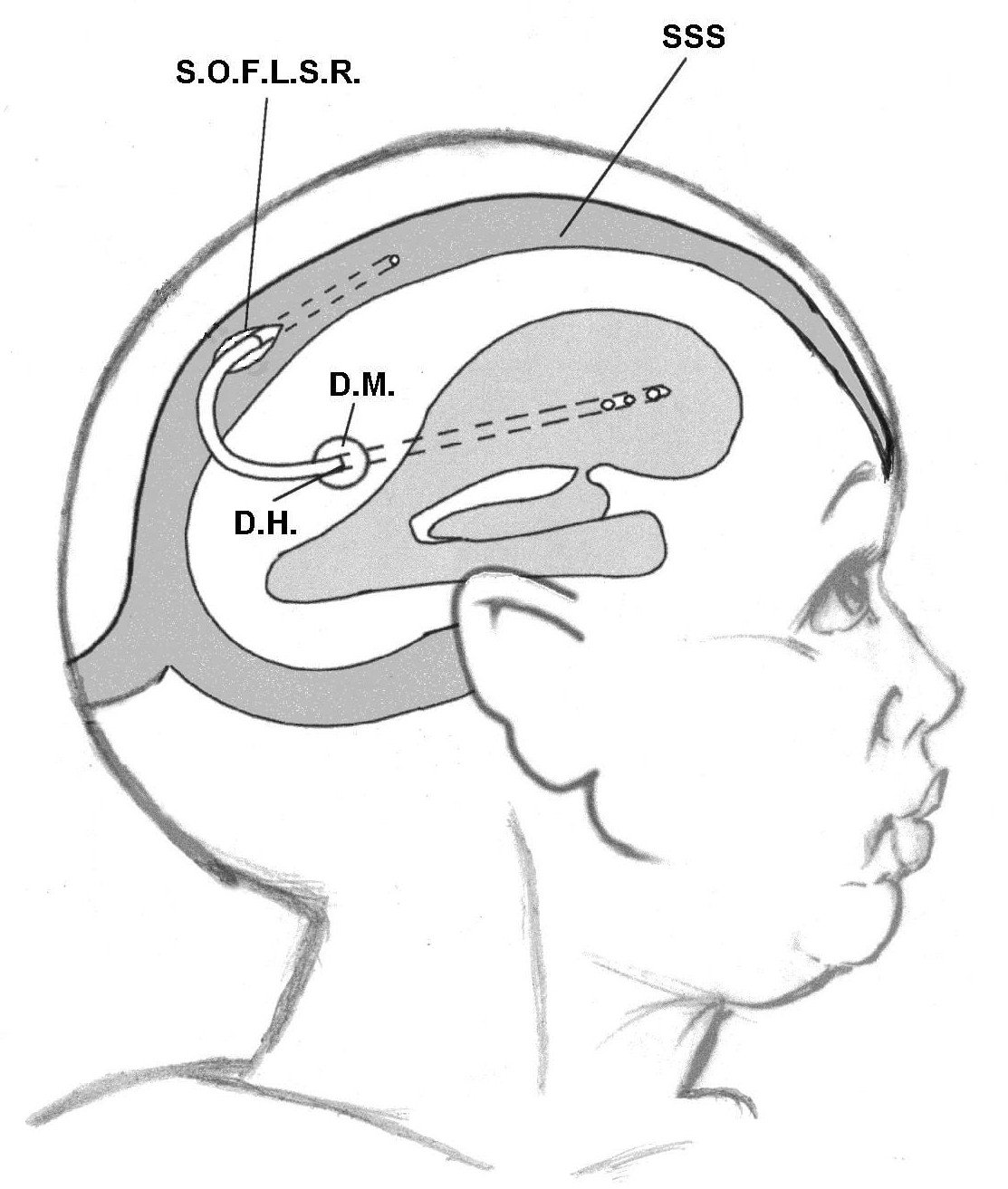
Definition
Hydrocephalus is a pathological condition characterized by the abnormal accumulation of cerebrospinal fluid (CSF) within the intracranial compartment. This accumulation leads to increased intracranial pressure (ICP) and results in a variety of neurological manifestations.
The existence of cerebrospinal fluid was known as early as the 18th century BC, as documented in the Edwin Smith Papyrus. Hydrocephalus itself was recognized by Hippocrates (Breasted, 1930).
From a pathophysiological perspective, hydrocephalus is defined as an imbalance between the production and absorption of CSF of sufficient magnitude to cause net accumulation of fluid within the cerebral ventricles or the intracranial cavity, leading to increased intracranial pressure (Milhorat, 1996).
Cerebrospinal Fluid Physiology
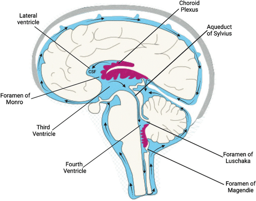
CSF is produced mainly by the choroid plexus, which is located:
- In the floor of the lateral ventricles
- In the roof of the third and fourth ventricles
The CSF circulates through the ventricular system as follows:
- From the lateral ventricles
- Through the foramina of Monro to the third ventricle
- Through the aqueduct of Sylvius to the fourth ventricle
From the fourth ventricle, CSF exits the ventricular system through:
- The foramina of Luschka
- The foramen of Magendie
It then circulates within the subarachnoid space around the brain and spinal cord. Final absorption occurs at the arachnoid granulations located along the superior sagittal sinus.
Thus, CSF occupies two main compartments of the central nervous system:
- The cerebral ventricles
- The subarachnoid space
The primary site of CSF absorption is the arachnoid villi, which represent evaginations of the arachnoid membrane into the dural venous sinuses.
Pathophysiology of Hydrocephalus
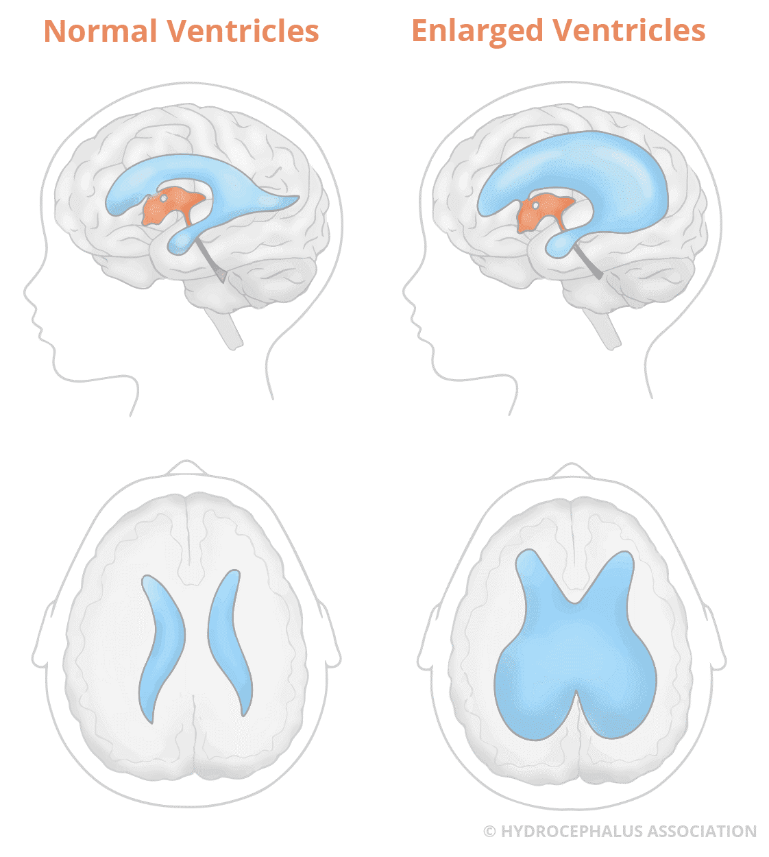
Because CSF is continuously produced and circulated, obstruction at any point along its pathway leads to:
- Proximal accumulation of CSF
- Increased CSF pressure
- Dilatation of the affected ventricular chambers
The resulting pathophysiological disturbances depend on:
- Patient age
- Skull expandability
- Duration of hydrocephalus
(Milhorat, 1996)
Types and Causes of Hydrocephalus
The overall incidence of hydrocephalus in the general population is unknown. However, the reported incidence of infantile hydrocephalus (3–4 per 1000 live births) is likely underestimated due to its association with many pediatric and adult intracranial disorders (Milhorat, 1978).
Classification Based on CSF Flow
Hydrocephalus is classified according to the level of CSF obstruction:
1. Obstructive (Non-Communicating) Hydrocephalus
Occurs when CSF flow is blocked within the ventricular system, preventing CSF from reaching the cerebral surface.
2. Non-Obstructive (Communicating) Hydrocephalus
Occurs when CSF reaches the cerebral surface, but absorption at the terminal drainage sites is impaired.
Etiological Mechanisms
Hydrocephalus may result from:
- Obstruction of CSF pathways
- Excessive CSF production
- Impaired venous drainage
Classification of Causes (Milhorat, 1972)
I. Obstructive (Non-Communicating) Hydrocephalus
A. Congenital Lesions
- Aqueductal stenosis:
- Gliosis
- Forking
- True narrowing
- Septum
- Atresia of the foramina of Luschka and Magendie (Dandy–Walker cyst)
- Intracranial masses:
- Benign cysts
- Vascular malformations
- Tumors
B. Acquired Lesions
- Aqueductal stenosis (gliosis)
- Ventricular inflammation and scarring
- Tumors and non-neoplastic masses
II. Non-Obstructive (Communicating) Hydrocephalus
A. Congenital Lesions
- Arnold–Chiari malformation
- Encephalocele
- Leptomeningeal inflammation
- Lissencephaly
B. Acquired Lesions
- Leptomeningeal inflammation:
- Infection
- Hemorrhage
- Particulate matter
- Intracranial masses
- Platybasia
C. Unproven or Doubtful Causes
- CSF overproduction
- Impaired venous absorption of CSF
Clinical Manifestations of Hydrocephalus
I. Infantile Hydrocephalus (Compressible Cranium)

Accurate history-taking is essential to differentiate congenital from acquired causes.
Key features include:
- Prenatal diagnosis by ultrasound
- Craniofacial disproportion
- Associated spinal defects (e.g., Chiari II malformation)
- Progressive head enlargement
- Bulging, tense anterior fontanel
- Dilated scalp veins
- Setting-sun sign (impaired upward gaze)
II. Adult Hydrocephalus (Rigid Cranium)
Four distinct clinical syndromes are recognized:
1. Acute Hydrocephalus
Characterized by rapid elevation of ICP leading to:
- Severe headache
- Vomiting
- Blurred vision due to papilledema
This condition is a neurosurgical emergency and may lead to brain herniation (coning). Additional signs include:
- Fever (in meningitis)
- Parinaud’s syndrome
- Diplopia due to sixth nerve palsy
- Altered consciousness, coma, or death
2. Chronic Hydrocephalus
Symptoms evolve gradually due to compensatory mechanisms and include:
- Mild dementia
- Behavioral changes
- Hormonal abnormalities from pituitary compression (hypopituitarism)
3. Normal Pressure Hydrocephalus (NPH)
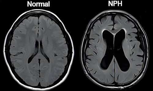
Defined as chronic hydrocephalus with normal CSF pressure but persistent ventricular enlargement.
Characterized by the classic triad:
- Dementia
- Gait disturbance
- Urinary incontinence
4. Arrested Hydrocephalus
A condition in which:
- CSF pressure has normalized
- No pressure gradient exists between ventricles and brain tissue
Investigations
- Fundoscopic examination to assess papilledema
- Cranial ultrasound in infants
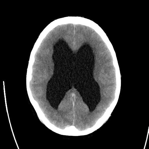
- CT scan to evaluate ventricular dilatation and periventricular edema
- MRI for detailed assessment of CSF pathways and intracranial masses
- Transcranial Doppler resistive index to assess ICP and shunt function
Treatment of Hydrocephalus
Medical Treatment
(Used as supportive or temporary therapy)
Surgical Treatment
- Removal of the obstructing lesion
The treatment of choice when feasible (e.g., tumors, cysts). - Reduction of CSF production
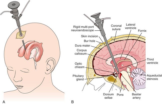
Choroid plexectomy (rarely used, endoscopic approach). - Bypassing the obstruction
Endoscopic Third Ventriculostomy (ETV).
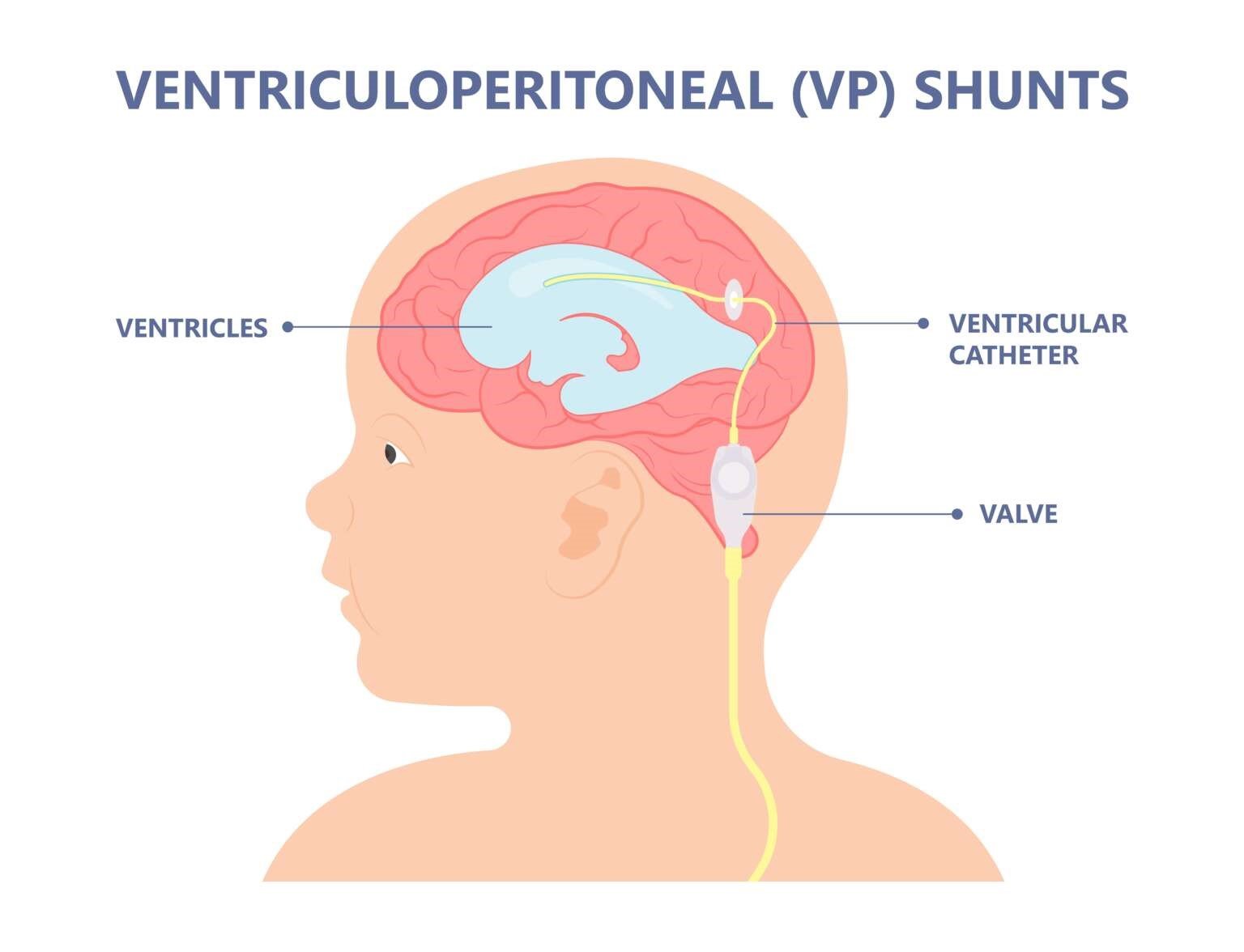
- CSF Diversion (Shunt Procedures)
- Ventriculo-peritoneal shunt
- Ventriculo-pleural shunt
- Ventriculo-atrial shunt
- Ventriculo-sinus shunt (El-Shafei RVS Shunt)